
Mesothelioma
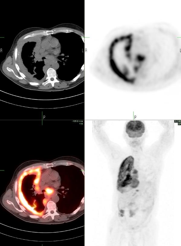
61 yrs old male presented with c/o productive cough for 3 months. He was initially treated with antibiotics for possible pneumonia, but without success. Subsequent chest CT found extensive pleural based soft tissue density encasing the right lung with possible mediastinal and diaphragmatic invasion, suspicious for mesothelioma.
PET-CT images show intense linear FDG uptake surrounding the right lung and extending into the right hemidiaphragm, corresponding to the soft tissue density on CT, max SUV measuring 9.5, consistent with mesothelioma. Multiple hypermetabolic nodes seen in the mediastium and the upper abdomen are consistent with metastatic disease.

1. The role of positron emission tomography/computed tomography in the diagnosis of pleural diseases. Orki A, Akin O, Tasci AE, Ciftci H, Urek S, Falay O, Kutlu CA Thorac Cardiovasc Surg. 2009 Jun;57(4):217-21.
2. 18F-fluoro-2-deoxy-D-glucose positron emission tomography and positron emission tomography/computed tomography imaging of malignant pleural mesothelioma. Subramaniam RM, Wilcox B, Aubry MC, Jett J, Peller PJ. J Med Imaging Radiat Oncol. 2009 Apr;53(2):160-9
3. Morphologic and functional imaging of malignant pleural mesothelioma. Yamamuro M, Gerbaudo VH, Gill RR, Jacobson FL, Sugarbaker DJ, Hatabu H Eur J Radiol. 2007 Dec;64(3):356-66.
This case was compiled by Dr. David He (BCM) and Dr. Joji Varghese (MEDVAMC)