PET-CT Imaging
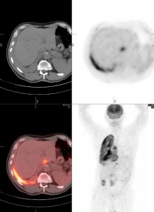
PET-CT is new and highly advanced imaging technique, which combines PET (positron emission tomography) and CT (computed tomography) in one machine.
The scanner merges PET and CT images together to provide a unique combination of both functional and structural information. It is very useful for detecting cancer, how far it has spread, to decide which treatment modality is best for you, and to monitor response to therapy. Brain and heart disorders are also studied using PET-CT scans.
 PET scanner creates an image of your body’s metabolism of a radio-active (or radio-tracer) material called FDG (F18-fluorodeoxyglucose), which is similar to glucose. For the scan you will receive an intravenous (IV) injection of small amount of FDG. Cancer cells metabolize glucose (and FDG) at a higher rate and PET scan shows this as abnormal activity.
A CT scanner uses x-rays to make an image of the sections of your body. The scan shows your body’s organs, bones and tissues in greater detail. For the CT scan, you will receive a contrast agent intravenously and orally. These help produce even clearer image.
When both PET and CT scanning are performed simultaneously, the information about function and structure are integrated through computer models to give a great deal of detailed information.
Fasting for a minimum of six to eight (6-8) hours (preferably overnight) is absolutely essential. In addition, a low carbohydrate (bread, potatoes, pasta, beans, rice, sugar, sweets) and high protein (meat, milk products, cheese, nuts, Atkin’s) diet the day before imaging helps in improving the image quality.
Prepare a list, which includes: names of medications you are currently taking, brief medical history, treatments you have had (Chemotherapy, Radiation Therapy, Surgery). Download the required forms, fill them to the best of your knowledge, and bring them with you. You should plan to be at the facility for 2-3 hours on exam day.
If you had other imaging studies performed such as CT scans, MRI, ultrasound etc, it is very crucial that the films and reports are made available to the radiologist who is interpreting the PET/CT scan. These films and reports will be returned to you or your referring physician, after review.
You are encouraged to drink 3-4 glasses of water prior to arrival for your test. Inform the staff performing the test if: you have ever had a reaction to a contrast agent or iodine in the past, if you have asthma, if you have a fear of small, enclosed spaces (claustrophobia). The actual scan takes approximately 35 – 45 minutes lying flat within the scanner. It would be best to come without any body jewelry.
Medications, other than for management of diabetes can be taken, as long as they are not required to be taken with meals. You should avoid vigorous physical exercise the day before, and on the day of the scan, and until the scan is completed.
If you are diabetic, you should take small, regular meals to keep glucose under control and continue to take your oral glucose medications or insulin. Please arrange the timing of the scan such that it is performed at least 4 hours after your last insulin injection. Patients with diabetes are often scheduled for the examination in the afternoon, thereby allowing them to take their morning diabetes medications and breakfast.
Confirm your appointment 24 hours prior to the test. Because of the extremely high demand for a PET/CT scan, your slot may be given to another patient.
The entire PET-CT scan takes about a total of two-three hours to complete. The staff will take a small blood sugar sample and place an intravenous line (IV) for a simple injection of the radioactive tracer (FDG).
The placement of the IV line will be slightly painful, but there should be no other pain involved. You will feel no side effects from the radioactive injection. You will also receive two bottles of oral contrast agent to drink.
No anesthesia is used. In some instances, if you have a history of anxiety in small, enclosed spaces (claustrophobia), you may be given a light sedative to help calm your anxiety and help you relax.
Following the injection, you will be asked to sit quietly for 45-60 minutes. This allows the drug to distribute in your body. You will then be positioned on a table for the actual scan. You will receive another injection of enhancing agent (used for the CT portion of the scan). You may notice warmth and flushing when the tracer is injected—this is normal. If you are allergic to either of the substances injected, you could have an allergic reaction. This can be treated with antihistamines and/or steroid medications. Rarely, an individual with already-existing kidney disease may experience further kidney damage.
You will then be imaged on the PET-CT scanner for approximately 30-40 minutes. The table will move slowly through a doughnut-shaped ring. You will need to lie still while the images are being taken. Later on, the technologist will process the scan and the images are made available for the radiologist to read. Every PET-CT scan is reviewed and correlated by both a board certified Nuclear Medicine Physician and a board certified Radiologist. Test results will be forwarded to your physician within 1-2 days.
You may go home when the study is finished and resume your regular diet and medication. You are encouraged to drink water to help clear the radioactivity from your body. If you have received sedation during the course of the exam, you will need someone to drive you home from the exam.
